
Blood Flow Restriction (BFR) for ACL Rehab & Post-Operative Care
Recovering from Anterior Cruciate Ligament Reconstruction (ACLR) or major orthopaedic surgery triggers a "perfect storm" for muscle loss:
Rapid Atrophy: Significant loss of muscle cross-sectional area (CSA) occurs within weeks post-surgery.
Load Constraints: Pain, joint effusion, and healing tissues limit heavy-load resistance training (70% 1RM)
Deficit Paradox: Muscle strength deficits can reach up to 40%, directly impacting long-term recovery and function.
At Ion Performance & Wellness, we utilize Blood Flow Restriction (BFR) therapy as an evidence-based bridge to restore muscle mass and strength without stressing healing tissues.
What BFR Is (and Isn't)
Originally developed in Japan as "KAATSU" training, BFR combines partial arterial inflow restriction with near-complete venous outflow restriction.
The Mechanism: Performing low-load exercise (20–40% 1RM) under vascular occlusion traps blood and metabolic byproducts in the working muscle.
The Clinical Role: BFR serves as an essential bridge for load-compromised patients—it is not a permanent replacement for heavy strength training once the joint and tissues mature.
Physiology of BFR
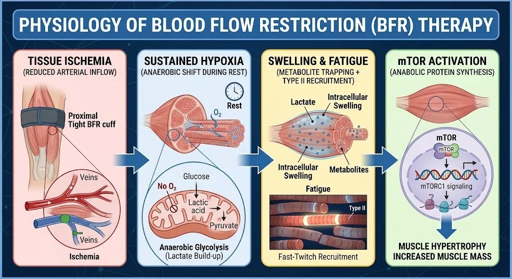
BFR Physiology summarized
Vascular Restriction: Partial arterial inflow is reduced while venous outflow is completely blocked.
Sustained Hypoxia: Oxygen deprivation shifts local muscle into anaerobic metabolism; keeping the cuff inflated during rest periods maintains this hypoxic environment.
Early Type II Recruitment: Metabolite buildup and intracellular swelling rapidly fatigue slow-twitch fibers, recruiting high-threshold fast-twitch (Type II) fibers at light loads.
Anabolic Signaling: Osmoreceptors detect fluid shifts, activating the mTORC1 pathway to stimulate muscle protein synthesis.
Evidence Summary
• Neuromuscular: Preserves quad mass & peak torque
• Symptoms: Reduces knee pain & joint effusion
• Functional: Improves IKDC & Lysholm scores
• RTS: Accelerates time to clear testing
Strength & Pain Relief: Hughes et al. (2019) demonstrated that low-load BFR (30% 1RM) matched heavy-load (70% 1RM) hypertrophy gains while achieving a 67% pain reduction (vs. 39% in controls) and superior effusion control.
Preserving Mass & Speeding RTS: Jack et al. (2023) showed BFR attenuated lean mass and bone density loss, reducing mean RTS functional clearance time to 6.42 months vs. 8.30 months in traditional rehab controls.
Acute Hypoalgesia: BFR provides immediate exercise-induced pain reduction, helping decrease arthrogenic muscle inhibition (AMI).
Expanding BFR: Applications Across Other Post-Operative Surgeries
While widely researched in ACL rehabilitation, BFR therapy is equally transformative across various post-operative lower and upper extremity surgeries where load tolerance is compromised:
Total Knee Arthroplasty (TKA) & Total Hip Arthroplasty (THA): Mitigates post-surgical muscular atrophy, accelerates gait mechanics recovery, and helps patients rebuild functional independence faster without overloading joint prostheses.
Rotator Cuff Repair & Shoulder Arthroscopy: Allows early low-load, high-metabolic activation of the deltoid and rotator cuff musculature while protecting delicate tendon-to-bone repair sites from excessive tensile stress.
Achilles Tendon Repair: Prevents rapid gastrocnemius and soleus atrophy during early immobilization or partial weight-bearing phases using passive BFR cycles or light active ankle movements.
Patellofemoral & Meniscal Repairs: Enables targeted quadriceps hypertrophy work during post-op range-of-motion or weight-bearing restriction phases, avoiding excessive patellofemoral compressive force.
Passive BFR
Protocol example (Takarada 2000, ACLR): 5 cycles of 5 min occlusion + 3 min reperfusion, 2×/day for 10 days.
Used to attenuate early thigh CSA loss when exercise is minimal.
Add cuff at individualized %LOP while using NMES. Neural + metabolic input
Goal: increase metabolic stress + fatigue → better hypertrophy stimulus.
Safety & Screening
Because venous thromboembolism (VTE) risk increases up to 100-fold in the first 6 weeks post-surgery, thorough clinical screening is non-negotiable:
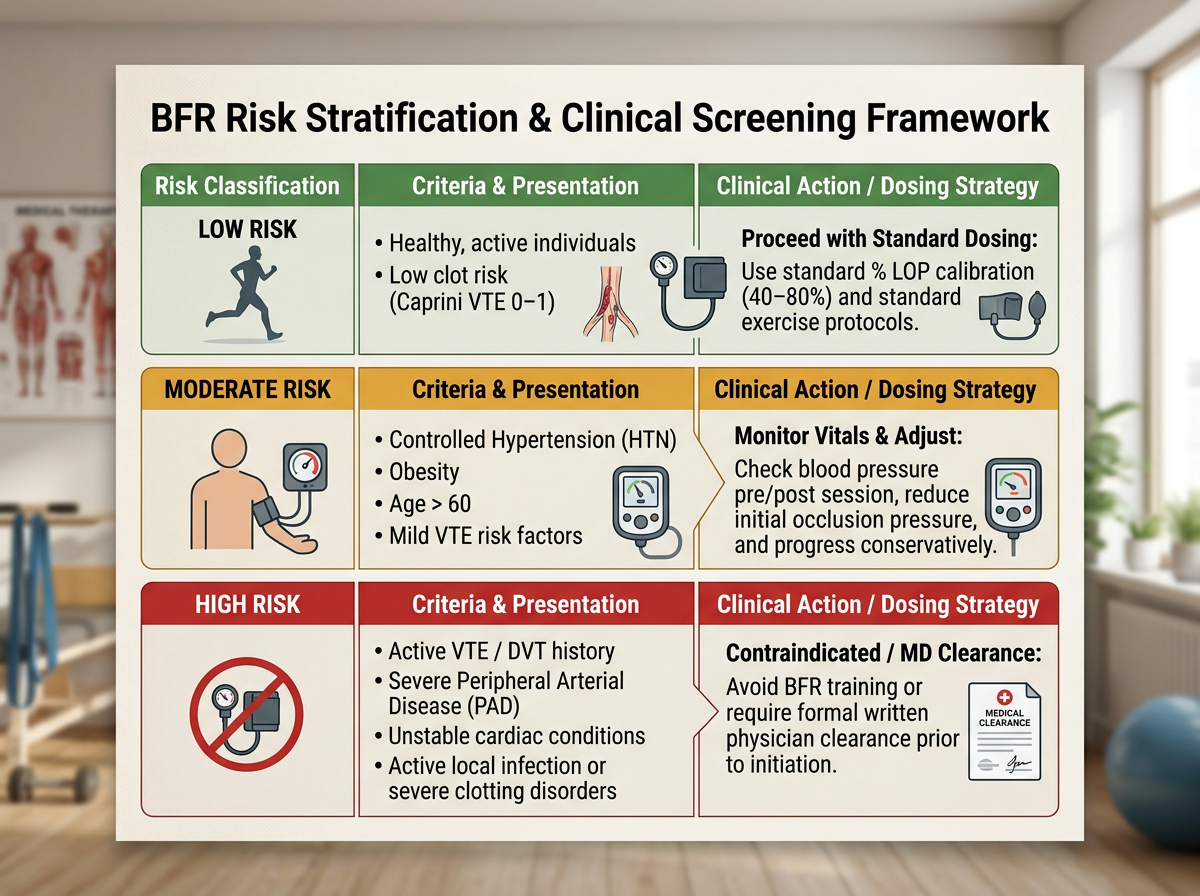
BFR Screening/Risk assessment
Absolute Contraindications: Active DVT/PE, severe Peripheral Arterial Disease (PAD), unstable cardiac conditions, active local infection, or severe clotting disorders.
Session Stop Rules: Immediately deflate the cuff if experiencing distal numbness, abnormal dizziness, skin color changes, or pain exceeding baseline levels.
Dosing Protocol & Sample Programming
Pressure: Calibrated to 40–80% Limb Occlusion Pressure (LOP) based on individual vascular measurement—never guess fixed pressures.
Load & Volume: 20–40% 1RM utilizing the standard 30-15-15-15 rep scheme (75 total reps) with 30-second inter-set rest periods.
Inflation: Keep the cuff inflated continuously throughout the 4-set exercise block; deflate between distinct exercises.
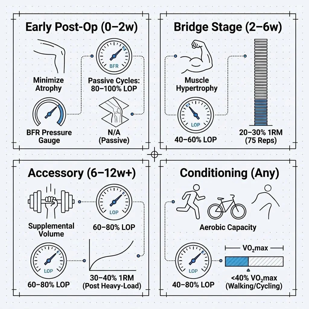
Sample BFR programming
Clinical Pitfalls to Avoid
Guessing Pressure: Always measure individual LOP/AOP via automated sensors or Doppler ultrasound.
Deflating Between Sets: Releasing pressure during 30-second rest intervals flushes out target metabolites and reduces the hypertrophic stimulus.
Delaying Heavy Load: Transition to heavy loading (>70% 1RM) as soon as joint capacity, effusion, and tissue healing allow.
Rebuild Strength Safer & Faster
Integrating BFR into post-operative rehabilitation allows our physiotherapy team to protect healing tissues while aggressively rebuilding muscle mass and functional capacity.
Book Your Post-Op Assessment with our clinical team at Ion Performance & Wellness in Burnaby today!